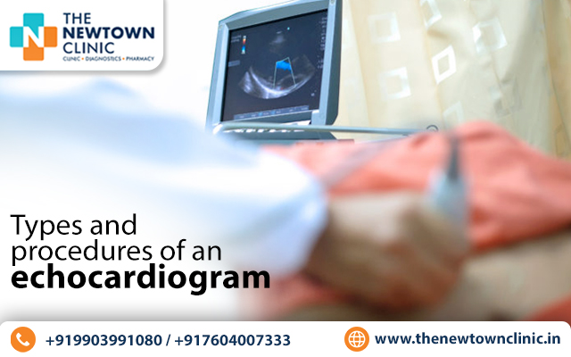
Echocardiogram is a non-invasive imaging technique that uses high-frequency sound waves to create a visual representation of the heart’s structure and function. They are used to diagnose and monitor various heart conditions, such as heart valve disorders, heart failure, and congenital heart defects. There are several types and procedures of echocardiogram that are used depending on the patient’s condition, says a cardiologist in Newtown.
Types of Echocardiogram
Below, a heart specialist in Newtown describes the different types of echocardiograms.
1. Transthoracic Echocardiogram (TTE): It is one of the most common procedures of echocardiogram and is performed by placing a transducer on the chest. The sound waves are transmitted through the chest wall, and the resulting images are displayed on a monitor. TTE is a painless procedure that takes around 30-45 minutes to complete.
2. Transesophageal Echocardiogram (TEE): This type of echocardiogram is performed by inserting a flexible tube with a transducer into the esophagus. This allows for a closer look at the heart’s structures and is particularly useful in diagnosing heart valve disorders and blood clots. TEE is performed under sedation and takes around 30-45 minutes to complete.
3. Stress Echocardiogram: This type of echocardiogram is performed while the patient is exercising. If the patient is unable to exercise, they are given medication that makes their heart beat faster, similar to when they exercise. As per the doctors of the best heart clinic in Newtown, it allows for a closer examination of the heart’s function under stress. The procedure can take up to an hour to complete.
4. Fetal Echocardiogram: This type of echocardiogram is performed on a developing fetus to detect any heart abnormalities. This procedure is performed by placing a transducer on the mother’s abdomen, and the images are displayed on a monitor. Fetal echocardiograms are typically performed during the second trimester and take around 30-45 minutes to complete.
Procedures of Echocardiogram
The top cardiologist in Newtown lists the different procedures of echocardiogram.
1. 2D Echocardiogram: This procedure captures two-dimensional images of the heart’s structures and functions. The images are displayed on a monitor, and the sonographer can examine the heart’s size, shape, and movement.
2. Doppler Echocardiogram: This procedure uses the Doppler effect to measure the speed and direction of blood flow through the heart’s chambers and valves. This allows the sonographer to detect any abnormalities in blood flow, such as blockages or leaks.
3. 3D Echocardiogram: This procedure captures three-dimensional images of the heart’s structures and function. This allows for a more detailed examination of the heart’s structures, particularly in complex cases.
Echocardiogram is an essential diagnostic tool in the management of various heart conditions. In case you have any concerns about your heart health, consult a cardiologist in Newtown, who may recommend an echocardiogram to help diagnose or monitor your condition.